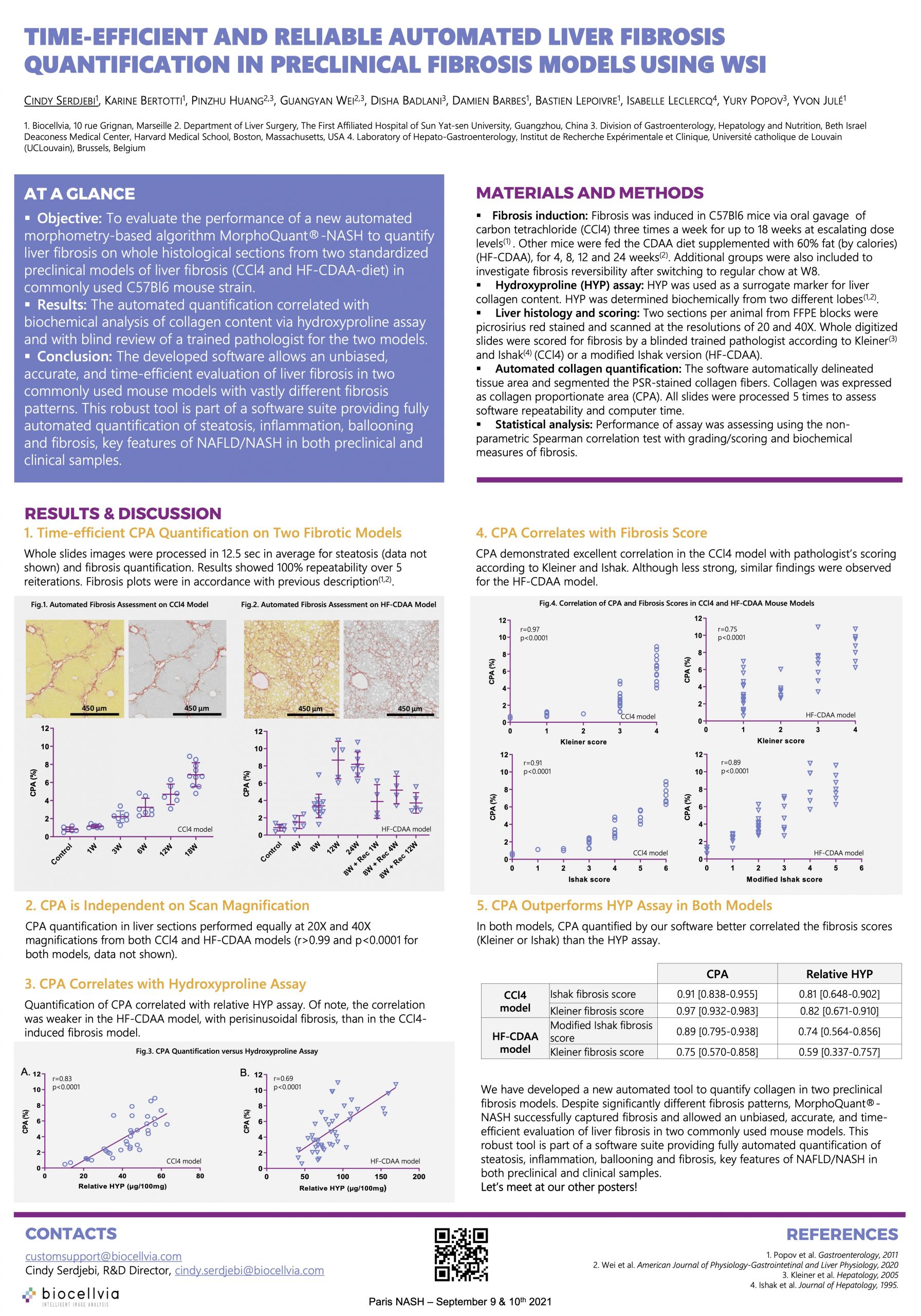
Objectives:
The aim of this study was to evaluate the performance of our automated morphometry-based algorithm MorphoQuant-NASH to quantify liver fibrosis on whole histological slide from two standardized preclinical mouse models of liver disease.
Methods:
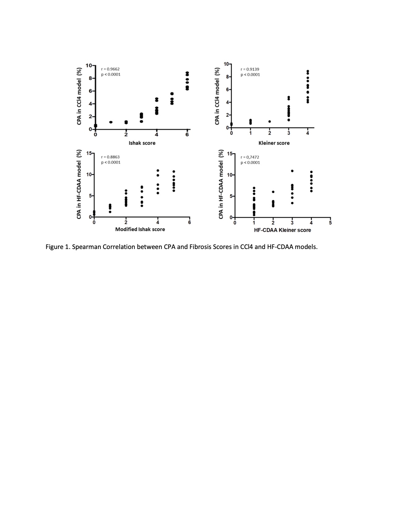
The chronic carbon tetrachloride (CCl4, predominantly septal fibrosis) and the high-fat choline-deficient amino acid (HF-CDAA)-diet feeding model of fibrotic steatohepatitis (predominantly perisinusoidal fibrosis) were assessed. Liver sections were stained with picrosirius red and whole slides were acquired at 20X and 40X optical magnifications. Automated analysis of collagen proportionate area (CPA) was performed on whole slide images, and expressed as the percent area occupied by all red collagen fibers versus the total section area. Biochemical analysis of relative hydroxyproline content was conducted and all sections were scored by an expert pathologist (IL) in a blinded manner according to Kleiner and Ishak (CCl4) or a modified Ishak (HF-CDAA). Performance of assay was assessed using Spearman correlation test with grading/scoring and biochemical measures of fibrosis. P-values < 0.05 were considered significant.
Results:
Fully automated quantification of collagen on whole liver slides took in average 12.5 seconds per section and results showed 100% repeatability over 5 reiterations.
CPA quantification in liver sections from both CCl4 and HF-CDAA models performed equally at 20X or 40X magnifications (20X vs 40X correlation, p<0.0001; r=0.9978 and 0.9984, respectively for each model). CPA measured by our software strongly correlated with hepatic collagen, determined biochemically (p<0.0001; r=0.8291 and 0.6877 for CCl4 and HF-CDAA, respectively). In the CCl4 model, CPA demonstrated excellent correlation with pathologist’s scoring according to Kleiner and Ishak (p<0.0001; r=0.9139 and 0.9662 for Kleiner and Ishak, respectively) (Figure 1). In HF-CDAA model, CPA showed strong correlation with the fibrosis scores (p<0.0001, r=0.7472 and 0.8863 for Kleiner and modified Ishak, respectively).
Conclusion:
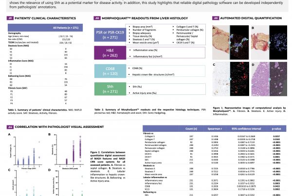
We have developed a software MorphoQuant-NASH that allows unbiased, accurate and time-efficient evaluation of liver fibrosis in two commonly used preclinical mouse models with vastly different fibrosis patterns. This robust tool is part of a software suite providing fully automated quantification of steatosis, inflammation, ballooning and fibrosis, key features of NAFLD/NASH, in preclinical and clinical samples.